


What it is: Magnetic Resonance Imaging (MRI) is one of the most advanced — and safest — ways to see inside the human body. Using a magnetic field and radio waves, it can help detect illness and injury in the bones, tissue and organs without the ionizing radiation used in conventional x-rays and CT scans.
How it works: MRI uses high power magnets and radiofrequency waves instead of x-rays to capture images to give physicians a literal view inside the body. MRIs produce soft tissue images and are used to distinguish normal healthy soft tissue from diseased or injured tissue. In some instances, an injection of contrast dye may be required.
Why it is used: This non-invasive technology is useful in diagnosing such things as multiple sclerosis, tumors of the pituitary gland and brain, strokes at their earliest stages, infections in the brain, spine or joints, and tendonitis. It is also used to visualize conditions related to sports injuries, and helps physicians evaluate masses in the soft tissues of the body, bone tumors, cysts, and bulging or herniated discs in the spine.
Preparing for an MRI Exam?
At Home
- Eat normally unless you have been instructed differently by your physician. Children and infants requiring sedation should not eat or drink approximately four hours prior to the MRI exam.
- Take any medication as usual.
- Bring a book, magazine, or something else to do while you wait for your exam.
At the MRI Center
- You will be asked your medical history.
- You will be told about your exam, then asked to sign a consent form.
- You will be asked to remove objects such as jewelry, hairpins, glasses, wig (if it has metal), hearing aids, and nonpermanent dentures. You will also empty your pockets of all objects.
- You may be asked to change into a hospital gown.
The MRI Exam
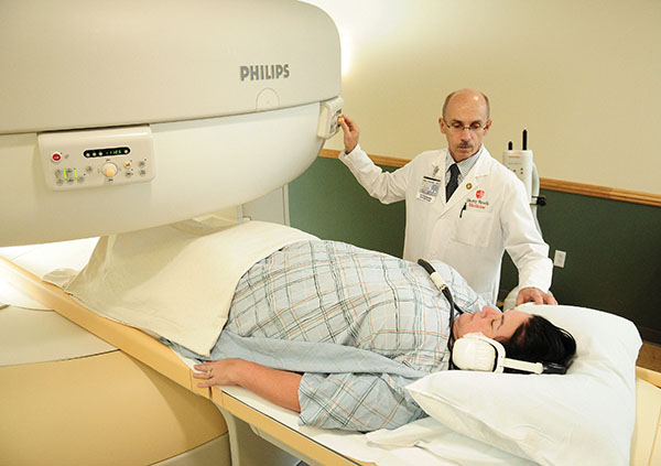
You will be positioned as comfortably as possible by the MRI technologist. The MRI examination table will slide into the magnet. The magnet may be fairly close to your face (within a few inches), depending on the type of exam you are having.
During the exam, you do not feel anything, but you will hear an intermittent knocking noise that may change in the frequency pattern of knocking. When the exam is finished, the images will be reviewed. If more images are needed, they will be obtained as soon as possible. You will be informed when you may leave the MRI center.
Restrictions
You will not be able to have an MRI examination if you have the following:
- Pacemakers
- Neurostimulators (Tens-Units)
- Ear implants
- Any metal clips in the eyes
Considerations
Please notify the MRI Center personnel and your physician if you are pregnant. Pregnant women may be examined by MRI in certain critical cases.
Fillings in your teeth, dental braces, and permanent bridges may cause some distortion of the MR image around the mouth area, but will not cause harm to you or the MRI equipment.
Total examination time usually ranges from 30 to 60 minutes.
It is very important that you hold very still for your MRI examination. If you feel this may be a problem, please inform your physician.
Different MRI Modalities at Stony Brook
At Stony Brook, we have multiple MRI machines so we can use the most appropriate technology for both the individual and the diagnosis. You will see MRI machines with a particular number designation such as 1T or 3T. The “T” refers to “Tesla,” which is the scientific unit that measures the power of magnets. The number designates how many Teslas the MRI machine uses. The higher the number, the stronger the magnet. Note, however, that the stronger magnet does not indicate a “better” machine. Different conditions may require different strength magnets for diagnosis.
1T Open MRI. In early 2012, Stony Brook Medicine acquired an open MRI machine. Although this technology has been available during the past decade, the image quality has not been comparable to that of traditional MRIs — until now, which is why Stony Brook has just acquired the technology. Currently, it is the only true open MRI in Suffolk County.
Why use an open MRI? With a traditional MRI, patients are sent slowly through a hollow, cylindrical structure in order to obtain a full body scan. For patients with claustrophobia or discomfort with enclosed or small spaces, the experience can be challenging. An open MRI in essence, revolves around the patient, eliminating the need to be in an enclosed space. The structure also makes it easier for patients to remain comfortable and still for the duration of the test. Other benefits:
- Ability to accommodate patient of large physical stature up to 500 to 550 pounds as well as patients with wide shoulders who may not fit traditional machines.
- Ideal for wheelchair-bound patients. With the open MRI, the bed on which the patient lies for the examination can move up and down automatically so a patient in a wheelchair can be easily and comfortably transferred to it.
- Ideal for children. The comfortable environment is also a good option for children and teens who otherwise might be too antsy for a traditional MRI. Parents can remain in the room, and even hold their child’s hand during the procedure. We also make music and videos available during the testing.
- Allows for additional specialty testing. This includes musculoskeletal, abdominal and neuro testing, as well as diffusion work and certain sequences — possible because the radiologists and MRI technologists can better position the patient to capture accurate images.
1.5T MRI. Stony Brook has two 1.5T magnetic resonance imaging scanners that offer increased speed, exceptional resolution and accuracy from earlier models. Long the gold standard for MRI imaging, 1.5T is ideal for diagnosing a wide range of medical conditions, performing complex MRI studies and helping physicians do surgical planning. The 1.5T scanner located in the hospital's new MRI Suite is wide bore, which is useful for imaging of larger patients or those who have mild claustrophobia.
3T MRI. This is an updated version of the MR scanner with the most powerful magnet available. It is particularly useful in diagnosing conditions involving the brain, spine and musculoskeletal system because it produces exceptional anatomic detail. It also produces images with increased spatial resolution, making it ideal for high-quality vascular imaging. Our newest 3T model, located in the new MRI Suite at the hospital, is wide bore, which is useful for imaging of larger patients or those who have mild claustrophobia.
If your physician has referred you to MRI testing, you can opt for either a traditional or open MRI at Stony Brook Medicine. The preparation is exactly the same.